 Add to Chrome
 Add to Firefox
 Add to Edge
Learning from Multiple Expert Annotators for Enhancing Anomaly Detection in Medical Image Analysis
Mar 20, 2022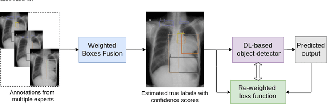
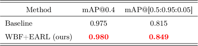

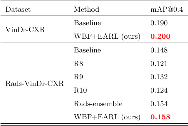
Building an accurate computer-aided diagnosis system based on data-driven approaches requires a large amount of high-quality labeled data. In medical imaging analysis, multiple expert annotators often produce subjective estimates about "ground truth labels" during the annotation process, depending on their expertise and experience. As a result, the labeled data may contain a variety of human biases with a high rate of disagreement among annotators, which significantly affect the performance of supervised machine learning algorithms. To tackle this challenge, we propose a simple yet effective approach to combine annotations from multiple radiology experts for training a deep learning-based detector that aims to detect abnormalities on medical scans. The proposed method first estimates the ground truth annotations and confidence scores of training examples. The estimated annotations and their scores are then used to train a deep learning detector with a re-weighted loss function to localize abnormal findings. We conduct an extensive experimental evaluation of the proposed approach on both simulated and real-world medical imaging datasets. The experimental results show that our approach significantly outperforms baseline approaches that do not consider the disagreements among annotators, including methods in which all of the noisy annotations are treated equally as ground truth and the ensemble of different models trained on different label sets provided separately by annotators.
Learning to Automatically Diagnose Multiple Diseases in Pediatric Chest Radiographs Using Deep Convolutional Neural Networks
Aug 14, 2021Chest radiograph (CXR) interpretation in pediatric patients is error-prone and requires a high level of understanding of radiologic expertise. Recently, deep convolutional neural networks (D-CNNs) have shown remarkable performance in interpreting CXR in adults. However, there is a lack of evidence indicating that D-CNNs can recognize accurately multiple lung pathologies from pediatric CXR scans. In particular, the development of diagnostic models for the detection of pediatric chest diseases faces significant challenges such as (i) lack of physician-annotated datasets and (ii) class imbalance problems. In this paper, we retrospectively collect a large dataset of 5,017 pediatric CXR scans, for which each is manually labeled by an experienced radiologist for the presence of 10 common pathologies. A D-CNN model is then trained on 3,550 annotated scans to classify multiple pediatric lung pathologies automatically. To address the high-class imbalance issue, we propose to modify and apply "Distribution-Balanced loss" for training D-CNNs which reshapes the standard Binary-Cross Entropy loss (BCE) to efficiently learn harder samples by down-weighting the loss assigned to the majority classes. On an independent test set of 777 studies, the proposed approach yields an area under the receiver operating characteristic (AUC) of 0.709 (95% CI, 0.690-0.729). The sensitivity, specificity, and F1-score at the cutoff value are 0.722 (0.694-0.750), 0.579 (0.563-0.595), and 0.389 (0.373-0.405), respectively. These results significantly outperform previous state-of-the-art methods on most of the target diseases. Moreover, our ablation studies validate the effectiveness of the proposed loss function compared to other standard losses, e.g., BCE and Focal Loss, for this learning task. Overall, we demonstrate the potential of D-CNNs in interpreting pediatric CXRs.
VinDr-RibCXR: A Benchmark Dataset for Automatic Segmentation and Labeling of Individual Ribs on Chest X-rays
Jul 03, 2021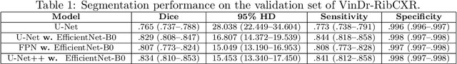
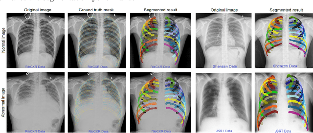
We introduce a new benchmark dataset, namely VinDr-RibCXR, for automatic segmentation and labeling of individual ribs from chest X-ray (CXR) scans. The VinDr-RibCXR contains 245 CXRs with corresponding ground truth annotations provided by human experts. A set of state-of-the-art segmentation models are trained on 196 images from the VinDr-RibCXR to segment and label 20 individual ribs. Our best performing model obtains a Dice score of 0.834 (95% CI, 0.810--0.853) on an independent test set of 49 images. Our study, therefore, serves as a proof of concept and baseline performance for future research.
Enhancing MRI Brain Tumor Segmentation with an Additional Classification Network
Sep 25, 2020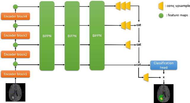
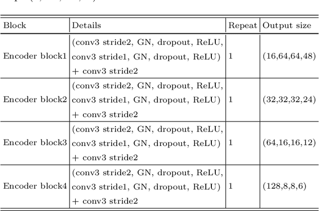
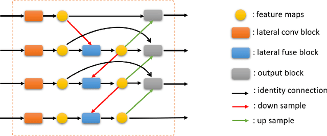
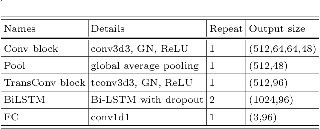
Brain tumor segmentation plays an essential role in medical image analysis. In recent studies, deep convolution neural networks (DCNNs) are extremely powerful to tackle tumor segmentation tasks. We propose in this paper a novel training method that enhances the segmentation results by adding an additional classification branch to the network. The whole network was trained end-to-end on the Multimodal Brain Tumor Segmentation Challenge (BraTS) 2020 training dataset. On the BraTS's validation set, it achieved an average Dice score of 78.43%, 89.99%, and 84.22% respectively for the enhancing tumor, the whole tumor, and the tumor core.
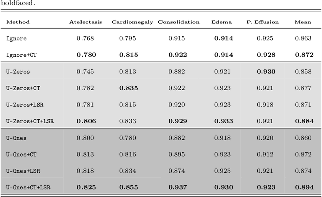
Interpreting Chest X-rays via CNNs that Exploit Hierarchical Disease Dependencies and Uncertainty Labels
May 25, 2020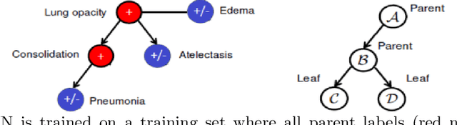
The chest X-rays (CXRs) is one of the views most commonly ordered by radiologists (NHS),which is critical for diagnosis of many different thoracic diseases. Accurately detecting thepresence of multiple diseases from CXRs is still a challenging task. We present a multi-labelclassification framework based on deep convolutional neural networks (CNNs) for diagnos-ing the presence of 14 common thoracic diseases and observations. Specifically, we trained astrong set of CNNs that exploit dependencies among abnormality labels and used the labelsmoothing regularization (LSR) for a better handling of uncertain samples. Our deep net-works were trained on over 200,000 CXRs of the recently released CheXpert dataset (Irvinandal., 2019) and the final model, which was an ensemble of the best performing networks,achieved a mean area under the curve (AUC) of 0.940 in predicting 5 selected pathologiesfrom the validation set. To the best of our knowledge, this is the highest AUC score yetreported to date. More importantly, the proposed method was also evaluated on an inde-pendent test set of the CheXpert competition, containing 500 CXR studies annotated by apanel of 5 experienced radiologists. The reported performance was on average better than2.6 out of 3 other individual radiologists with a mean AUC of 0.930, which had led to thecurrent state-of-the-art performance on the CheXpert test set.
Interpreting chest X-rays via CNNs that exploit disease dependencies and uncertainty labels
Nov 18, 2019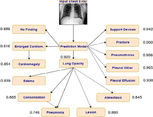
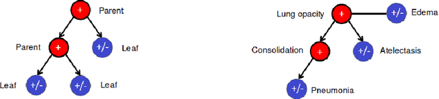
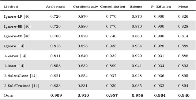
Chest radiography is one of the most common types of diagnostic radiology exams, which is critical for screening and diagnosis of many different thoracic diseases. Specialized algorithms have been developed to detect several specific pathologies such as lung nodule or lung cancer. However, accurately detecting the presence of multiple diseases from chest X-rays (CXRs) is still a challenging task. This paper presents a supervised multi-label classification framework based on deep convolutional neural networks (CNNs) for predicting the risk of 14 common thoracic diseases. We tackle this problem by training state-of-the-art CNNs that exploit dependencies among abnormality labels. We also propose to use the label smoothing technique for a better handling of uncertain samples, which occupy a significant portion of almost every CXR dataset. Our model is trained on over 200,000 CXRs of the recently released CheXpert dataset and achieves a mean area under the curve (AUC) of 0.940 in predicting 5 selected pathologies from the validation set. This is the highest AUC score yet reported to date. The proposed method is also evaluated on the independent test set of the CheXpert competition, which is composed of 500 CXR studies annotated by a panel of 5 experienced radiologists. The performance is on average better than 2.6 out of 3 other individual radiologists with a mean AUC of 0.930, which ranks first on the CheXpert leaderboard at the time of writing this paper.